How AI-powered segmentation and automated volumetric calculations are transforming the way radiologists measure, report, and act on imaging findings.
For decades, volumetric measurements in radiology meant manually tracing organ boundaries, recording dimensions slice by slice, and typing values into a report. It was painstaking, time-consuming, and — perhaps most critically — inconsistent. Two radiologists measuring the same structure on the same scan could arrive at meaningfully different numbers, introducing variability that undermined clinical confidence and complicated longitudinal tracking.
Today, artificial intelligence is replacing that entire process. AI automatically segments anatomical structures, calculates true three-dimensional volumes in seconds, and populates those results directly into structured reports — all before the radiologist opens the case. The measurement burden that once consumed minutes of every complex read has been quietly automated away, and the impact on workflow, accuracy, and patient care is substantial.
What Is Automated Segmentation in Radiology?
Segmentation is the process of delineating specific structures — tumors, organs, lesions, vascular territories — within a medical image. Traditionally, this required a radiologist or imaging technologist to manually outline boundaries on each individual slice, a process that could take anywhere from several minutes for a single lesion to considerably longer for complex, multi-structure examinations. The result was highly dependent on the individual performing the task, their experience, fatigue level, and the ambiguity of the image itself.
AI-based segmentation replaces this with deep learning models — particularly convolutional neural networks (CNNs) — trained on large datasets of expert-annotated images. Once trained, these models can identify and outline structures across CT, MRI, and PET/CT scans with remarkable speed and consistency. They do not tire, do not rush, and apply the same learned pattern recognition whether they are processing the first case of the day or the hundredth. Research has demonstrated that AI segmentation reduces inter-reader variability from over 15% to under 8%, depending on the measurement standard applied — a clinically meaningful improvement for oncology follow-up, surgical planning, and treatment response assessment.
Volumetric Calculations: From Slices to 3D in Seconds
Once a structure is segmented, volumetric calculation follows automatically. This is a significant advance over the two-dimensional diameter measurements that have long dominated radiology reporting. RECIST criteria, which govern tumor response assessment in oncology trials, rely on the longest diameter of a target lesion — a practical compromise designed for the era of manual measurement, not a true reflection of three-dimensional disease burden. AI removes that compromise entirely, computing true 3D volumes from the full imaging dataset without additional effort.
The clinical significance is substantial. Volumetric measurements are more sensitive to early changes in tumor burden, particularly for lesions that are irregular in shape or respond asymmetrically to treatment. Early detection of progression — or response — can directly influence treatment decisions, potentially sparing patients from ineffective therapies sooner. Tools such as TotalSegmentator can segment over 100 anatomical structures in a single CT scan simultaneously, generating volume and attenuation measurements across the liver, spleen, kidneys, lungs, cardiac chambers, and beyond — results that previously required dedicated post-processing workstations and significant technologist investment.
Results as Overlays and New Series
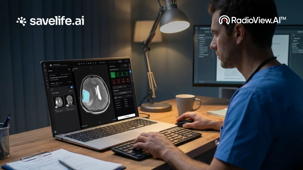
One of the most practical design choices in modern AI segmentation platforms is how results are delivered to the reading radiologist. Rather than generating a separate PDF or external output that must be opened and cross-referenced, AI measurements are presented directly within the PACS viewer — either as color-coded overlays on the original images or as a new image series that can be toggled alongside the source scan.
This integration keeps the radiologist firmly in their primary workflow. They can review the AI’s segmentation boundaries, verify that organ outlines are accurate, and make manual corrections in edge cases — all without switching applications or interrupting their read. This also maintains clinical accountability: the radiologist is not passively accepting AI output, but actively reviewing it as part of their interpretive process. The AI handles the repetitive, high-volume measurement task; the radiologist applies the clinical judgment that no algorithm can replicate.
Automated Population Into Structured Reports
The final and perhaps most transformative step in this workflow is the seamless transfer of AI-derived measurements into the radiology report itself. Using DICOM Structured Reporting (DICOM-SR) and standardized Common Data Elements (CDEs), AI systems can auto-populate structured report templates with organ volumes, lesion dimensions, and attenuation values at the moment of reporting. The radiologist no longer types these figures — they review pre-filled values, confirm their accuracy, and sign off.
This approach has been validated in real clinical environments. AI systems have successfully segmented and auto-reported liver and spleen measurements across thousands of CT examinations, with values integrated directly into the body of the report. Beyond time savings, auto-population enforces documentation completeness — ensuring that key measurements are always included, always formatted consistently, and always traceable back to the AI model that generated them. The downstream effect is cleaner data for follow-up tracking, fewer omissions in oncology reporting, and more reliable communication with referring clinicians.
The Clinical Impact: Speed, Consistency, and Better Patient Care
The cumulative effect of automated segmentation, three-dimensional volumetrics, and structured report auto-population is a radiology workflow that is fundamentally faster and more reliable than its manual predecessor. Turnaround times decrease as the measurement burden is offloaded to AI. Diagnostic consistency improves as human variability is reduced. Reports become richer with quantitative data that supports better clinical decision-making across multidisciplinary teams.
In high-volume departments managing hundreds of studies per day, these gains compound quickly. Saving three to five minutes per complex case across a full shift reclaims hours of daily capacity — time that can be redirected toward complex interpretation, patient communication, and multidisciplinary collaboration rather than data entry. It also directly addresses one of the most pressing challenges facing radiology today: the growing imbalance between rising imaging volumes and a constrained radiologist workforce. Some AI-assisted reporting environments have demonstrated efficiency improvements of up to 40%, a figure that translates directly into reduced backlogs, faster diagnoses, and better outcomes for patients.
RadioView.AI™: Fast, Accurate AI Measurements Built for Clinical Trust
RadioView.AI™ is purpose-built to deliver fast, accurate AI Measurements that integrate directly into radiology reporting workflows. Its automated segmentation and volumetric tools give radiologists instant, quantitative results they can act on immediately — eliminating manual measurement steps, reducing turnaround times, and supporting more consistent, data-rich reports across every modality.
What sets RadioView.AI™ apart is not just the speed and accuracy of its measurements, but the clinical trust infrastructure built around them. In a healthcare environment where patient data security is non-negotiable, RadioView.AI™ is both HITRUST and HIPAA Compliant — meeting the most rigorous standards for healthcare data protection and privacy. Every measurement, every auto-populated report value, and every patient record processed through the platform is secured to the highest level the industry demands.
For radiology departments, imaging centers, and health systems evaluating AI measurement tools, RadioView.AI™ offers the combination that matters most: clinical-grade accuracy, seamless workflow integration, and enterprise-level compliance. It is not just a faster way to measure — it is a smarter, safer foundation for the future of volumetric reporting.
Conclusion
AI-powered segmentation and volumetric calculations represent one of the most impactful advances in radiology reporting in recent memory. By automating measurement, delivering results as visual overlays within the PACS environment, and populating structured reports without manual input, these tools remove significant friction from the imaging workflow. The outcome is faster reporting, more consistent measurements, and better-informed clinical decisions — achieved not by replacing radiologists, but by giving them capabilities that were previously impossible at scale. As platforms like RadioView.AI™ continue to advance AI Measurements with full HITRUST and HIPAA compliance, the new standard for volumetric reporting is not on the horizon. It is already here.
FAQs
1. What is AI segmentation in radiology?
AI segmentation uses deep learning models to automatically outline anatomical structures — such as organs, tumors, and lesions — within medical images. It replaces manual boundary tracing with consistent, reproducible measurements that reduce inter-reader variability and accelerate the reporting process.
2. How accurate are AI volumetric measurements compared to manual measurements?
Studies show that AI-driven segmentation reduces measurement variability significantly, with inter-reader variation coefficients dropping from over 15% with manual tracing to under 8% with AI assistance. This level of consistency is particularly valuable in oncology, where accurate, reproducible measurements directly influence treatment decisions.
3. How do AI measurements appear in the radiology workflow?
AI results are delivered as color-coded overlays within the PACS viewer or as a new imaging series alongside the original scan. This allows radiologists to review, verify, and if needed correct segmentation boundaries without leaving their primary reading environment.
4. Can AI measurements be automatically added to radiology reports?
Yes. Using DICOM Structured Reporting (DICOM-SR) and standardized Common Data Elements (CDEs), AI-derived organ volumes, lesion dimensions, and attenuation values are auto-populated directly into structured report templates — reducing manual data entry and improving documentation consistency.
5. Is RadioView.AI™ compliant with healthcare data security standards?
Yes. RadioView.AI™ is both HITRUST and HIPAA Compliant, meeting the highest standards for healthcare data security and patient privacy. Every measurement and patient record processed through the platform is handled with enterprise-level data protection.
6. Why is volumetric measurement more valuable than diameter-based measurement?
Diameter measurements under RECIST criteria reflect only one dimension of a lesion, which can miss early changes in irregularly shaped or asymmetrically responding tumors. True 3D volumetric calculations capture the full extent of disease burden, providing a more sensitive and clinically meaningful measure of treatment response.